Karl Whittington • University of California at Berkeley
Recommended citation: Karl Whittington, “The Cruciform Womb: Process, Symbol and Salvation in Bodleian Library MS. Ashmole 399,” Different Visions: New Perspectives on Medieval Art 1 (2008). https://doi.org/10.61302/GLRT6998.
Introduction
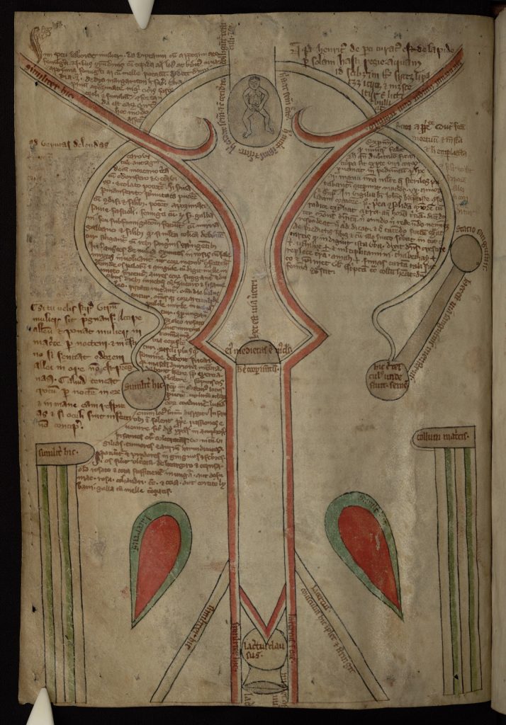
Among the medical texts and illustrations that make up MS Ashmole 399 in the Bodleian Library in Oxford lies an image of striking graphic power and beauty (Figure 1). Colored lines curve and twist, connecting abstract shapes and irregular fields of text. At the top corners of the manuscript page, two red lines curve down towards the center, stopping abruptly and jutting out to form two points before continuing as parallel straight lines to the bottom of the page. Around them, shapes lie across the largely symmetrical surface: two black lines arch over the top of the red lines, connecting to two spheres that float near the center of the page. In the bottom corners, two columnar forms anchor the composition, and just inside of them lie two large teardrop-shaped red forms, outlined in green ink. At the top stands a tiny human, enclosed in a shaded oval. Small captions and labels cover parts of each shape, while longer texts weave haphazardly around them. This image is a diagram of the female sexual anatomy, from a thirteenth-century book of medical texts and illustrations. Modern viewers can decipher easily only a few of these forms: to us, the drawing resembles some kind of map or abstract diagram more than a representation of actual anatomy, or anything else recognizable, for that matter. Strangest of all to our eyes, there is no contour or outline of the external body in this representation.

Fig. 1. Female anatomy, Oxford, Bodleian Library MS Ashmole 399, fol. 13v.
The only visible body is the small figure at the top. The forms describe a system unconnected to and seemingly independent of the body as a whole. While this is the case with anatomical diagrams, where the tiniest parts of the body are blown up to stand on their own, this particular image provides an extreme example – its forms are completely two-dimensional, and exist without concrete boundaries or obvious connections to a larger system. The texts and labels that penetrate the diagram’s space and engrave themselves on the organs only enhance its bizarre non-bodily look. Through these labels, however, we can identify the diagram’s main features: the fetus, womb, fallopian tubes and ovaries, cervix, vaginal canal, vaginal muscles, and “stations” for menstrual blood: what we see then, are the essential features of the medieval understanding of female reproductive physiology, mapped out on a page.
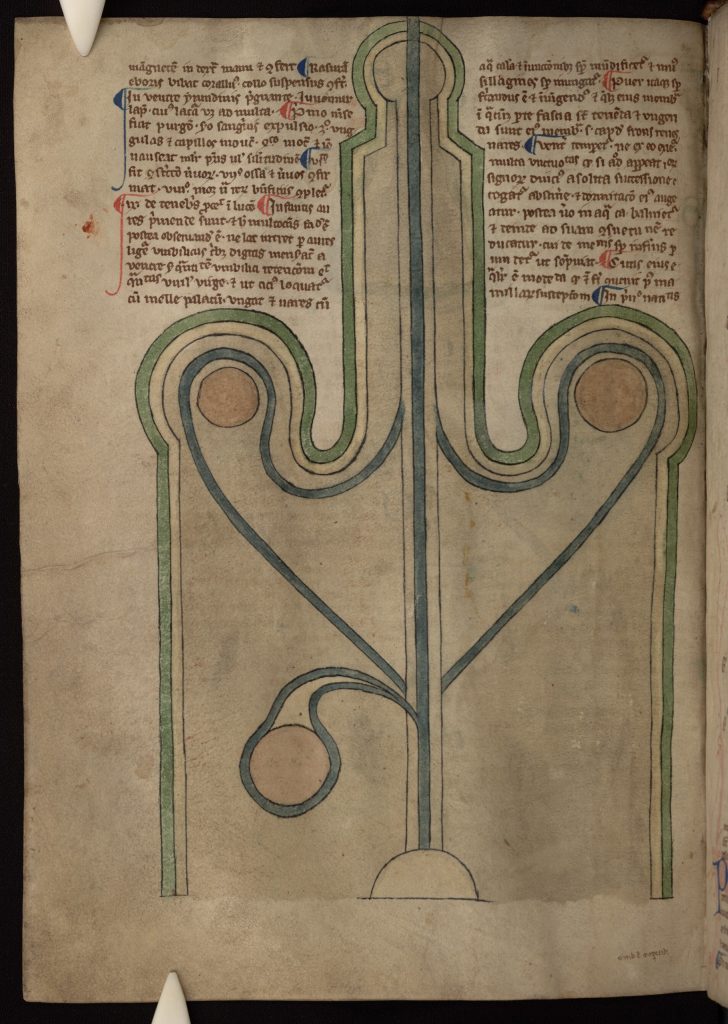
This diagram, and the accompanying image of the male genitalia (Figure 2), is well known in the literature on medieval anatomy and also in studies of medieval gender construction, medicine and generation theories. Among the many commentators, Monica Green, Danielle Jacquart, and Claude Thomassett, especially, have dealt specifically with this image of the female anatomy, exploring its meaning in relation to medieval models of reproduction, gendered sexual and reproductive roles, women’s health issues, and the transmission of classical and Islamic medical knowledge into the west.[1]

Fig. 2. Male reproductive anatomy, Oxford, Bodleian Library MS Ashmole 399, fol. 24v.
However, scholars have not explored fully the diagram’s visual appearance, and how the specific appearance and orientation of the diagram’s forms change our understanding of the ways that medieval scientists conceptualized the reproductive female body as a site both of theoretical physiological processes and abstract systems, symbols and ideologies. This essay explores the diagram’s representational strategies, especially as they compare to other images in this manuscript. Examining the specific arrangement of its forms, I aim to address the flexibilities of medieval diagramming practices more generally, and to show that this enigmatic example actually draws on diverse subjects and traditions of representation to create one image that remained logical and cohesive to its original viewers. Rooting the formal elements of this “scientific” image within a broader context than is often attempted will lead us far from the conventional understanding of medieval anatomical images, but closer, I hope, to an understanding of the diverse meanings and implications which may have been intended by the image’s makers or received by its viewers.
To accomplish this integration of a medieval “medical” image into broader discourses of theology, contemporary image-making and gender, I will employ Madeline Caviness’s “triangulatory” approach to medieval images, in which both historical evidence and contemporary theoretical perspectives are brought to bear on the medieval object. In the more theoretical sections of my argument, I will be drawing mainly on the work of Caviness and Margaret Miles; these sections are not guided by a single theoretical model or position, but are more broadly influenced by the issues of viewing, physical orientation, and gender construction raised by these and other scholars. The historical side of my argument, in contrast, examines more closely the contemporary implications of the religious symbolism that I observe in the image, and seeks to explain how the drawing’s visual strategies speak to other types of medieval art, both diagrammatic and pictorial.
The Manuscript
MS Ashmole 399 consists of seventy-eight vellum folios, and contains over two- dozen medical texts on topics of physiology, obstetrics, and generation, as well as several series of illustrations.[2] This type of book is often referred to as a “medical miscellany,” and it stored a hodgepodge of medical knowledge, rather than a specific program of texts and images. The illustrations were completed in the 1280s or 1290s in England, and the texts were likely filled in during the subsequent few decades. When and in what order the texts were written on and around the images remains an open, and likely unanswerable question; my analysis of the manuscript, however, suggests that its scribes may have planned to include both texts and images from the start, and spaced the image-cycle accordingly.[3] The readership of such a manuscript would likely have consisted of physicians, mathematicians, and natural scientists, and though we know nothing of the manuscript’s specific provenance- it may have been made for a university, monastery, or an independent individual patron- it clearly participated in established scientific discourses.[4]
The manuscript’s texts include diverse authors. Most numerous are medical tracts by Constantine the African, a Muslim drug merchant who, following his conversion to Catholicism and while in residence at the monastery at Monte Cassino, was a primary agent in the transmission into the west in the eleventh and twelfth centuries of Muslim (and thus Classical) medical traditions.[5] Many of the illustrations in the Ashmole manuscript belong to an ancient, possibly Roman type of anatomical illustration known in the modern literature as the Fünfbilderserie, which was actually a series of nine drawings, not five, of the major bodily systems.[6] Fragments of this series, first pieced together by Karl Sudhoff around 1900, appear throughout late-Antique and medieval medical manuscripts, though the full series of nine drawings occurs only in four surviving manuscripts. The Fünfbilderserie was strictly a series of drawings, not texts, and thus the texts in Ashmole 399 are not necessarily meant to explain the images; here, text and image transmit related but separate bodies of knowledge.
One artist or workshop likely completed most of the drawings in MS Ashmole 399; there are seventeen distinct pages of illustration, all of which use the same four color- washes (blue, green, red and yellow/tan) and similar uses of line and shading with dark brown ink.[7] Folios 33 and 34 are the only exceptions: now perhaps the best known images in the manuscript, they depict a “case history” of a doctor and a female patient, and were likely inserted at a later date.[8] Monica Green terms the cumulative effect of text and image in the manuscript, “a veritable summa on generation,” and it betrays a sustained interest in female reproduction.[9] Still, the manuscript deals, for the most part with physiology rather than practical medicine: the images are meant to show how processes of the body work, and in some cases to describe their physical appearance and nature in addition to their mechanical function. In the late thirteenth century, most practical medical issues relating to the female reproductive system and childbirth were the domain of trained female midwives, whose learning came from a separate tradition of practical medical knowledge that was primarily transmitted orally or written in the vernacular.
Reproductive Process and Physical Orientation
Turning to the diagrams of the male and female genitalia, we must ask not only how the forms of the body are arranged and placed, but also how they place the reader through the physical orientation on the page. This is one of the key approaches that has not yet been explored in relation to these kinds of anatomical drawings: it is important to ask how the drawings position their makers and viewers in terms of physical space and, correspondingly, gender. As we will see, both drawings are operating for and from a specifically male viewpoint.
While the image of the female anatomy confuses the viewer in the arrangement of its forms, the image of the male reproductive anatomy exhibits symmetry and overall visual clarity. Unlike in the diagram of the female genitalia, the image requires no labels to explain itself to the assumed male viewer; rather, he presumably could recognize the forms by their relation to their physical referents. The male image, in other words, operates more as a description of natural forms, instead of as an explanation of theories or processes, the model through which I will argue the female image functions.[10] The penis and testicles dominate the page, rendered almost architecturally in their solidity and clarity of form. With the help of medieval medical sources, including Constantine the African’s De Spermate and De Coitu within the Ashmole manuscript, one may trace the path of the semen, represented by the thick lines that start from the bottom of the page, branch off to circle around the testicles, and continue upward and outward through the urethra.[11] The round object in the lower left corner, the only element breaking the image’s symmetry, most likely represents the bladder, or possibly the prostate.
The male diagram’s interior cross-section viewpoint constructs a hieratic image, juxtaposing the visual dominance and solidity of the forms with the upward movement and propulsion of the dark lines, shaded in green and blue. Most significantly, however, the image orients the male genitalia towards the presumed male viewer, its base contiguous with the reader’s own body, the organ itself seen from above and as if in an x-ray. I would also suggest that the penis and testicles in this image are really the only parts of the external body in all the drawings in MS Ashmole 399 that are approximately “life-sized” – that is to say, it is approximately the size of an average adult male erect penis (in the manuscript, it measures about 5.5’’ by 1.5’’ inches), again relating the image directly to the body of a male viewer, further placing him as the image’s “subject.” The drawing of the female anatomy, however, reverses this visual orientation, as if in order to maintain the perspective of a male viewer. Here, the reader views the female organs from an outside vantage, facing the body, rather than from the subjective orientation adopted for the male diagram. Seen from this perspective, the female system seems to have no boundaries: there is no containing edge or outline between the bodily organs and the outside world, or the body and the text.
Though as symmetrical as the male-anatomy image, the female diagram does not embody the same kind of visual unity. The main organizing principle is the pair of continuous red lines that divide the page, defining, from bottom to top, the walls of the vagina, the cervix, and the upper walls of the womb. Near the top, offshoots from these lines proceed upward into the womb, suggesting a bicornuate effect, the two “horns” of the womb; this interior septum divides the womb and the body into two parts.[12] Arching over the top of these red lines are two parallel gray lines, which appear to signify the fallopian tubes, ending in the spherical ovaries. At the bottom of the page we see the outer view of the vaginal opening and, on either side of it, two teardrop-shaped objects, which in turn are flanked by two columnar shapes.
When viewed in connection with Constantine the African’s De genecia, included in MS Ashmole 399, and other contemporary gynecological texts, and when analyzed with the text of the labels written onto the organs in the drawing itself, certain features of the illustration make more sense while others become more confusing. The two teardrop- shaped organs, colored red, are each labeled lacertus (muscle), as are several of the other small shapes near the entrance to the vagina at the bottom center of the manuscript page; presumably these point out the system of muscles that controls the physical process of orgasm and childbirth. The text of Constantine the African’s De genecia also describes one of the orifices of the uterus, the collum matricis (neck of the womb), in which coitus was said to occur; this corresponds in fol. 13v to the two column-like shapes at the lower edges, one of which is indeed labeled collum matricis and the other similiter hic.[13] According to Charles Singer, these represent a bisected vagina, thus the “entrance” to the womb described by Constantine and others.[14] His De genecia also speaks of certain “pits” in the main chamber of the uterus (also identified as collum matricis) from which the menstrual flow originates.[15] The circular organ connected to the left ovary, labeled statio sanguinis (station of the blood), seems to correspond to one of the “pits” described by Constantine.
In medieval sources generally, and in this diagram specifically, there is an overall ambiguity about the terminology of the uterus. The entire diagram, which is usually described as the uterus and adnexa, its accompanying parts, bears little relation to the images of the fetus in the womb on the following page, which look like upside-down vessels or vases. These correspond only to the small illustration at the top of the uterus, on fol. 13v, the human figure enclosed in an oval; but there the womb is certainly more than just the oval membrane around the fetus. Similar confusion surrounds the phrase collum matricis, which describes an organ and also an entrance. This confusion on our part in understanding what words such as uterus or collum matricis meant in the thirteenth century reflects, I believe, confusion and inconsistency among medieval anatomists on precisely the same issues. The uterus was an organ, but also a system; the collum matricis an entrance, but also a main chamber.
In his close analysis of the diagram’s labels, Charles Singer argues that the image actually describes how the reproductive system changes in its two different states: pregnant and non-pregnant.[16] Following his argument, the dark red lines delineate the boundaries of the womb when the woman is not pregnant, while the lightly shaded lines that curve downward show its boundaries during a pregnancy (as well as illustrating the path of female sperm through the fallopian tubes). The diagram includes two female “testicles,” or ovaries, and alludes to two more, off to the sides in the upper corners, where the labels point out the path of female sperm to the non-pregnant uterus. The details of this double-representation are confusing, but the importance lies in the fact that it depicts two systems at once, using the visual tools of shading and color in addition to the labels. The forms present themselves not as individual bodily organs, but as explanatory signs, many carrying multiple identifications, constituting a highly elastic representational system. Again, we see that the explanation of processes (intercourse, conception and pregnancy) is more important than the description of a physical body, contrasting with the diagram of the male genitalia, which depicts the forms of the body as static and changeless – continually erect and ejaculating.
Text and Image
University-trained physicians in the mid to late thirteenth century had access to an extensive body of classical literature on theories of reproduction, most notably in the works of Galen and Aristotle, who offered two very different ways of thinking about the body.[17] In the Aristotelian model, male and female are polarized and given different physical values and conditions to a far greater degree than in earlier, Hippocratic medicine; men are defined by warmth, and women by coldness, which reinforced other medieval binaries of superiority and inferiority, ability and inability, and activity and passivity.[18] Most importantly and controversially, Aristotle insisted that women did not produce sperm: rather than playing an active reproductive role, they provided only the vessel and nourishment for the male seed. Integrated into his larger natural-philosophical system, this notion of one-sided conception would be greatly influential throughout the Middle Ages. It was transmitted especially through Constantine the African’s translation of Avicenna’s Canon of Medicine, itself based on Aristotle, reinforcing a pattern of setting the sexes in opposition through an emphasis on their difference.
Galen, however, argued that both the male and female testes produced sperm, which came together inside the womb to form the fetus, thus de-emphasizing the difference between the sexes in the reproductive process.[19] He focused on the physical similarities between male and female genitalia, arguing that both possess testes and produce sperm, and that the physical structure of their sexual organs were similar, though the female’s are interiorized. This way of framing female anatomy, as equivalent to the male’s but interiorized (hidden and therefore inferior), would certainly be considered sexist by today’s standards, but it does give the woman equal participation in the creation of human life.
Though it is difficult to place the Ashmole diagrams into a specific dialogue with these two contemporary models of medieval reproduction, they seem overall to bear a greater correspondence to Galen’s reproductive theories than they do to Aristotle’s. In the diagram of the female reproductive system, the vagina really does resemble the phallus of the male diagram, interiorized, and the woman’s large and prominent ovaries allude to the similarly sized and shaped male testicles.[20] Certainly, many of Galen’s ideas correspond to Aristotelian notions of female passivity and inferiority, but the diagrams seem to betray an interest, as do many of Constantine the African’s texts, in rendering Aristotelian theories of female passivity compatible with influential traditions of medicine that acknowledged the presence of female seed- specifically, the new medical ideas, influenced by Galen, emerging around this time from the Muslim world.[21]
Despite the difficulty of establishing precise text-image connections in MS Ashmole 399, the ways that the texts interact physically with the images on the page remains an unexplored and productive path to follow. The main texts penetrating the image from the upper corners detail recipes for conception and aiding childbirth, and thus certainly relate to the diagram, but do not explain it: that job belongs to the labels written on the diagram in a different hand. The text of the labels is consistent with other labeling texts throughout the manuscript, and seems to have been the original and earlier one, while the invasive texts on the outside are most likely later, and initiate a new discourse on treatment and herbal medicine. These texts in the margins sneak around the corners of the various parts of the female anatomy, weaving in and out between the pregnant and non-pregnant wombs, paying no attention to the boundaries that are so insistently respected in the male diagram, in which two neat columns of text lie in the top corners of the page. In the female diagram, the discourses of science and medicine are inscribed on, in and around the spaces of the female “body,” both in the original labels and explanations and in the later texts that creep in from the margins.
Perhaps even more significant is the absence of a real “body” in the representation, made even more clear by the two-dimensionality of these texts covering the diagram. Madeline Caviness has written extensively about the ways in which medieval representations denied women real bodies, instead replacing women with metaphors or fetishes.[22] As in Caviness’s description of Mary’s “disemboweling” in the viewer’s entrance into the architectural space of the medieval-church-as-Mary, the haphazard inscription of male science onto the diagram’s forms disembodies the female “figure,” turning a physical body into an abstract system. The inscription of male science onto the female reproductive system, as well as the assumed orientation towards the male viewer discussed earlier, is analogous to the male gaze which, as Caviness and others argue, denies the represented female wholeness or agency.[23] The later texts inserted on the image represent what is perhaps an anxious attempt by scientists to demystify the “secrets of women” that the diagram represents.[24] And while some of the text on the page does in fact aim to be helpful to women, in giving pharmaceutical recipes to lessen the pain of childbirth, the way that it is placed on the page still displaces the female body, and, in the process, also displaces the physicality of the experience of childbirth.[25]

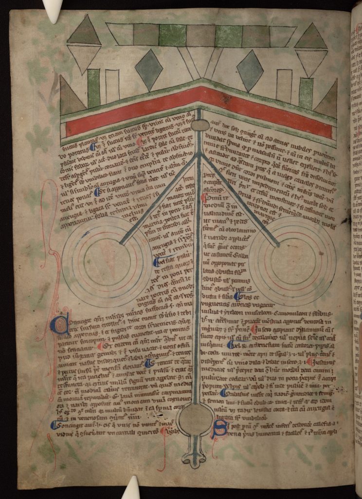
Fig. 3. Eyes and brain, Oxford, Bodleian Library MS Ashmole 399, fol. 22v.
Perhaps significantly, the only other illustration in the manuscript in which the text encroaches on the body’s space is the diagram of the eyes and the brain, suggesting a possible connection between the two, as organs which were understood to be fundamental sites for the reception of outside stimuli (Figure 3).[26] In this diagram, highly abstract forms depict the two eyes, the nose, and presumably the nerves carrying sensory information to the brain, which is strikingly represented by the abstract blocks of color at the top of the page. Susannah Biernoff has recently explored the medieval relationship between sight and reproductive flesh, arguing that vision, in the High Middle Ages, carried with it varying degrees of carnality – she describes “ocular desire” (affective responses arising from the physical pleasures of sight), “wounding gazes,” and other medieval metaphors connecting sight with the rest of the body.[27] These two diagrams in the Ashmole manuscript may demonstrate how such ideas of physical seeing and ocular desire appear in diverse forms of imagery: the drawings depict both the female genitalia, a medieval site of carnality, and the eyes and brain, home to the senses, as sites of transmission, reception, and passing through – they visualize the process of this transmission, rather than depicting carnality directly, in the form of a body. Thus, as processes, they require labels or text within their forms, although it is important to stress that the text in the diagram of the eyes and brain, like the text surrounding the female diagram, does not “explain” the images per se. I do not wish to argue a strict correspondence between ocular desire and sexual desire within these images, but rather to point out that there are structural and formal similarities between the systems of sight and reproduction in the manuscript, to which other medieval sources and metaphors testify.
These two images are also the only two in the manuscript that describe their subjects conceptually, rather than perceptually – the drawings of the male genitalia, skeleton, heart, or viscera, for example, adhere visually to their natural physical referents at least in part; though they were likely not the result of direct observation of human or animal subjects, their mode of illustration still aimed to depict and describe natural forms. The female anatomy and the brain diagrams, however, reveal theoretical diagrams of things unseen; in the case of the female diagram, it renders visible the “secrets of women,” secrets which were apparently still in need of explanation even for this manuscript’s presumed academic audience.[28]
Situating the Cross
The issue of things “unseen” leads to perhaps my most unexpected reading of this image: the evocation, within the forms of the female anatomy, of the shape of the crucified Christ. Although hitherto “unseen” or un-remarked in the expansive secondary literature on this image, the suggested figure is inescapable once noticed. The oval-enclosed fetus at the top of the page doubles as Christ’s head, while the curving lines running down the center of the page that define the boundaries of the womb, cervix and vagina also evoke the contours of two arms and a proportional, if slightly elongated body; their red color isolates them from the surrounding forms, making the suggestion of a body more pronounced. The two “horns” of the uterus are elongated and provide a neck leading up to the head. At the center of the page, the red lines protruding outward near the cervix call to mind representations of Christ’s loincloth, and as the lines run toward the upper corners, they evoke the graceful curve of the arms so common in English imagery of the crucified Christ during the thirteenth century.

Fig. 4. Arma Christi, Passional of Abbess Kunigunde, Prague National Library MS XIV A 17, fol. 3v. Photo by Zde; CC-BY-SA-4.0.
The image resembles not only the form of medieval crucifixes, but also images of the arma Christi, or instruments of Christ’s Passion.[29] Moving outwards from the evoked body, the fallopian tubes and circular ovaries imply the form of the flail used to beat Christ at the scourging, complete with a handle (the statio sanguinis, or “blood station” mentioned above). The two shapes in the bottom corners, labeled collum matricis, look a great deal like actual columns, such as the one Christ was tied to while being whipped. Most evocative, however, are the two tear-drop lacerti that so closely resemble medieval representations of Christ’s side-wound. A slightly later illumination of the arma Christi from Prague (Figure 4). clearly illustrates this unmistakable visual similarity: the shape and two-toned shading of the side wound in the Prague illustration are nearly identical to the “muscles” in the Ashmole diagram.[30] It is also interesting to note, besides their visual similarities, the conceptual correspondence between the extensive medieval tradition of arma Christi representation and the Ashmole illustration of the female anatomy: both explain processes through an emphasis on the individual elements. By taking elements out of the narrative or process, both image-types call attention to the part-by-part explanation of a larger story or system. In the case of the arma Christi, Passion devotion was reduced to meditation on individual moments; in the Ashmole female diagram, symbolic power resides visually within individual organs, separate from their role in the overall system.
Although this suggestion of crucifixion iconography within a reproductive diagram may seem bizarre and incongruous, a number of medieval precedents for this image provide visual, textual, and theoretical frames for understanding its meaning. Other medieval diagramming traditions, especially cartography, use the cross in a similar way- as an organizing principle for viewing that which, like the inside of the female body, was unseen and un-seeable to medieval viewers.[31] On a basic level, most medieval world maps organize the continents themselves into the shape of a cross, through the “tripartite” mapping system, also sometimes called the “T-O” map type. The thirteenth-century German map from Ebstorf (destroyed in 1943) crystallizes this cruciform re-organization explicitly, depicting Christ’s head, hands, and feet extending from the corners of the map; this incorporation of the space of the earth into Christ’s body turns the whole world into a macrocosm of Christ’s saving flesh. The application of this motif in the Ashmole drawing may carry this salvific message as well, pointing to the redemptive power of birth and reproduction in general, or perhaps even alluding to Christ’s assuming human flesh or Mary’s role in Christ’s entry into the world.
In addition to their similar cruciform organization, maps were also used as explanatory tools for processes.[32] Daniel Connolly has recently written about the use of maps as step-by-step tools for “imagined pilgrimages,” mental journeys through the maps, which he argues were an established part of devotional practice in England in the twelfth and thirteenth centuries. In these diverse examples, we can see medieval English cartographic traditions working in similar ways to anatomical drawings, prioritizing processes over exact forms, while rendering their forms legible through symbolic frameworks.
This use of the cross as an organizational principle in medieval graphic design is a topic requiring further research, but it is appealing to point to the connections between the anatomical and cartographic traditions in southern England in the thirteenth century as evidence of common goals and representational strategies. Besides these cartographic comparisons, other approaches might likewise “explain” the cruciform re-organization of the Ashmole image. One could certainly argue for a mnemonic function – that the image of the crucifix and the arma Christi would have been so familiar to any medieval viewer that matching up its various components to different parts of the medical image would have been a powerful mnemonic tool for remembering the shapes and relative positions of the parts of the female reproductive system. In an entirely different vein, we could even see the inclusion of the cross as a way of “masculinizing” the female body. Though these possibilities are intriguing, my aim in the final section of this essay is not to attempt to prove in any sense that the cruciform elements of the image were intentional and constructed (though this may indeed have been the case). Rather, what interest me most are the possible frames through which viewers may have seen this cruciform reorganization of the body’s forms, which, whether intentional or subconscious, reveal the cross’s flexibility as a symbol within this image’s pictorial and conceptual system. Before engaging this question, however, I want to address how we might locate the image within medieval habits of diagramming, and briefly consider how this diagram, which still appears extraordinary and even alien to the modern viewer, made logical sense within thirteenth-century representational practices.
In her recent book on the various modes of medieval visual communication, Jean Givens proposes a theoretical model for analyzing the representational techniques of natural-scientific and botanical drawings that proves useful when dealing with anatomical images such as those in the Ashmole manuscript. She outlines three categories for such drawings: they may be realistic, naturalistic, or descriptive (and an image may fall into more than one category).[33] She uses the term “realistic” to address images of things, the existence of which is not in question, “naturalistic” for images “that register the overall irregularity and variety inherent in living creatures,” and “descriptive” for images that intend to communicate visually something about the “external and sometimes internal physical structure of real-world objects and phenomena, (though) they need not be lifelike.”[34] The Ashmole female diagram is thus decidedly “descriptive”: it aims to communicate information about the inside of the female body, and also, as I now propose, about its symbolic connection to the crucifix or to salvation.
Ignoring the external appearance of its natural referent, and certainly not addressed by the term “lifelike,” the Ashmole diagram conveys information over appearance, and conceptual connections over natural form. Medieval natural-scientists were surely aware that the female reproductive system was not cruciform in its actual shape, but in a diagram of this system, its form was not what was being addressed, even though some may still characterize the image as “anatomical.” Just as in maps, a great deal of empirical knowledge concerning the actual appearance of forms did not preclude their reorganization under a unifying visual principle, in these cases the cross, and in this process the images emerged without losing their value of truth. An interesting comparison on this point is the “sketchbook” of Villard d’Honnecourt. These drawings display a similar combination of description and symbolic hierarchy, and highlight the presence of geometric order within natural objects – a “neo-Platonic Aristotelianism” of nature embodied through universals.[35] Here, we can at least begin to see the ways in which the Ashmole diagram’s forms conform to certain medieval diagramming and pictorial practices, rather than diverging from them in a radical way, as the recognition of the crucifix might first lead one to perceive. The revelation of internal logic through generalization, rather than particularization, and the lack of contradiction between reading “real” and “symbolic” forms into one image simultaneously, are characteristic rather than exceptional aspects of the Ashmole image’s formal presentation.
Christ’s Female Flesh
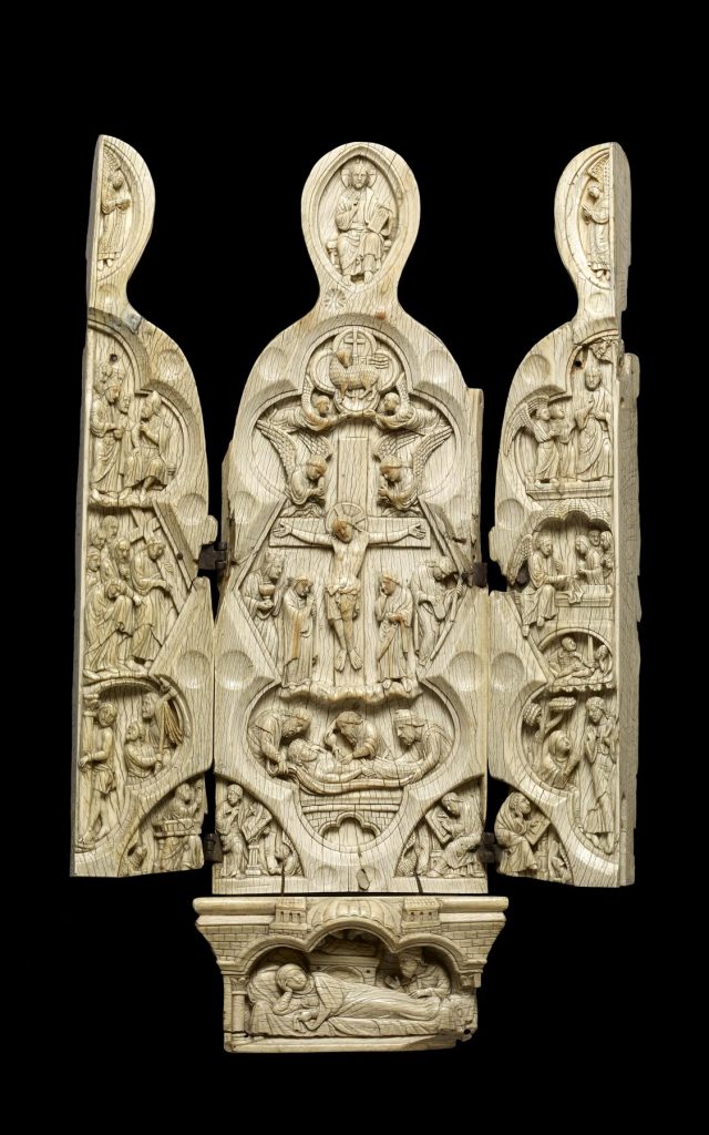
A comparative image quite far from these diagramming traditions in both function and execution, but no less striking in its conceptual similarity to the Ashmole diagrams, is offered by the figures known as Vierges ouvrantes, small sculptures of the Madonna and Child carved throughout Europe in the later Middle Ages that fold open to reveal a smaller sculpted image of Christ on the inside – usually either a crucifixion or a mercy-seat trinity. Figure 5 shows a 13th century French example from the Walters Museum in Baltimore.[36] Here, Christ’s crucified body is positioned at the center of the Virgin’s body, placed directly over the space where her womb would be, and, with relation to the female body, visually oriented in the same direction as the evoked crucifix in the Ashmole diagram. The conceptual correspondence between this image and the Ashmole diagram is evident: both reveal Christ’s crucified form within the female body, though certainly in very different ways, and for different audiences. Another fascinating comparison, in which the physical form of the cross is found within the female body, is the legend of St. Clare of Montefalco, the flesh of whose heart was said to bear an image of the cross when she was examined after her death.[37] All of these images suggest the achievement of salvation through enfleshing, specifically in female flesh.

Fig. 5. Vierge Ouvrante, French, Walters Museum, Baltimore.
The evocation of the crucified body in the Ashmole diagram, as literally cloaked in and composed of female flesh, emphasizes both the flesh and humanity that Christ took on at the incarnation, and also the flesh that he ultimately triumphed over in his death on the cross – indeed, the image could be read as a meditation on, or even theorization of the incarnation, exploring the meaning behind God’s enfleshing. Even more simply, it reinforces the idea that Christ entered the world through a woman. Caroline Bynum and others have explored how some twelfth and thirteenth-century writers emphasized the importance of the flesh as a path to God, rather than an obstacle, because of the shared humanity of women and Christ.[38] As Bynum writes, “the symbolic association of humanity with the female derived strength from the association of humanity with physicality (woman being the symbol of the flesh) and from the associations of Christ’s humanity with his mother.”[39] By the thirteenth century, men and women used Mary as a route to Christ in their ever-increasing desire to become closer to his flesh, in ocular and physical communion, and in meditation upon his physical body.[40]
It is too great a stretch to argue that the Ashmole diagram is meant specifically to depict Mary’s womb, with the Son of God contained therein, but viewers may have made this connection. Mary was often described in both visual and textual terms as, “the tabernacle, the vessel, container, the robe, and the clothing of Christ,” and the Ashmole diagram may depict this cloaking of Christ in the flesh received from his human mother.[41] The manner in which ordinary women related to Mary’s experience of the incarnation changed greatly over time, but by the thirteenth century writers emphasized more than ever Christ’s real birth from a real woman.[42] Margaret Miles argues that women’s lack of “religious subjectivity” (her term for religious self-determination in thought and action) outside of convents or beguine communities caused women like Mary, Eve, or Mary Magdalen to be simply figures within male discourse rather than religious subjects themselves. However, the thirteenth and fourteenth-century emphasis on Mary’s humanity may suggest otherwise – that women identified concretely with the Virgin’s experience of childbirth and motherhood.[43]
The Ashmole diagram’s medical aspects offer other connections between Christ’s body and women’s bodies even more directly. Spiritual trends in the thirteenth century often emphasized Christ’s role as nurturer and protector, and especially his role in feeding, through both body and blood.[44] The physiological connection between milk and menstrual blood that was at the core of medieval medical theory, which maintained that breast milk was the refined product of menstrual blood, which itself fed the fetus before birth, was not lost on artists, theologians or mystics.[45] Makers of images and texts referenced the connection countless times in order to demonstrate the significance of Christ’s blood as food and nourishment, referring to the wound in Christ’s side as a breast that suckles mankind, and as the passage through which he gave birth to his Church. A well-known French moralized bible shows just such a “church-birth,” in which the personification of the Church is literally born through Christ’s side wound.[46] The captions in the Ashmole diagram reference the movement of blood in great detail, identifying the via sanguinis menstrui, the statio sanguinis mentioned above, where the blood was produced and stored, and the specific place where the nourishing menstrual blood reaches the fetus, labeled his nutrit infans et crescit. Medieval devotional texts refer often to parallels between the redemptive possibility of Christ’s blood and women’s blood, but their visual juxtaposition in the Ashmole manuscript appears unique. Here, the woman’s blood that nourishes the infant becomes, visually and symbolically, Christ’s blood nourishing the soul.
Discussion of Christ’s and women’s blood raises in turn issues of birth and pain, especially recalling the “birth” of the church at the Crucifixion; medieval sources often describe Christ’s “labor” not in abstract terms, but rather with metaphors that are excruciatingly visceral. The Carthusian prioress Marguerite of Oignt provides the best- known example among modern scholars; near the end of the thirteenth century she wrote of Christ’s “church-birth,”
But when the time came for you to be delivered, your labor pains were so great that your holy sweat was like great drops of blood…Ah, Sweet Lord Jesus Christ, who ever saw a mother suffer such a birth! For when the hour of your delivery came you were placed on the hard bed of the cross … and your nerves and all of your veins were broken. And truly it is no surprise that your veins burst when in one day you gave birth to the whole world.[47]
This passage describes Christ’s physical and painful birth of his Church as being analogous to the pain of women in childbirth, just as the Ashmole diagram could be seen as juxtaposing the same two events, so similar in their painful physicality.
One may elaborate on this connection by comparing the image with another subject of frequent representation: Mary swooning at the side of the cross during the Crucifixion, experiencing the birth pains that, in medieval theology, were spared at Jesus’s birth but returned full-force at his death.[48] These images, which became common only in the fourteenth and fifteenth centuries, but nonetheless provide an interesting point of comparison with the Ashmole diagram, depict Mary in labor, her pain visually juxtaposed and rendered comparable to Christ’s, through body position and facial expression that draw the two experiences together. One way to view this comparison between Mary’s and Christ’s pain is to see it as one which ennobles the experience of childbirth through a comparison to divine suffering; as we have already seen, the Ashmole diagram’s medical emphasis concerns the reproductive system, through its piece-by-piece textual explanation of generation, and serves to highlight processes associated with childbearing, without illustrating it directly. The other side of the coin, however, might be to locate this juxtaposition in terms of an opposition, rather than a similarity. Medieval traditions consistently associated the pain of childbirth, as well as women’s flesh generally, with original sin; the pain of Christ’s death on the cross was the act that promised redemption from that sin, and an eventual escape from women’s pain that was tied to it so closely.[49]
In some ways, we are now far from the specific forms of the image that began this discussion, but I nonetheless believe that the image’s original viewers, monks or university doctors (both certainly trained in the exegesis of religious and philosophical texts and images), could have considered just these sorts of comparisons and correspondences. It’s impossible to know which, if any, of these many metaphors the diagram was primarily intended to catalyze, but the correspondences and range of visual associations that we find do attest to the flexibility of the image’s interpretation, shown both in the multiple anatomical elements embodied by the forms, and by the suggested crucifix image.[50] Perhaps ultimately we could say that the image offers the viewer a life cycle, with implied meanings and comparisons at each stage: the beginning, shown through the explained processes of conception and birth, the end, Crucifixion, and the ultimate rebirth of humanity through Christ’s saving death.
Conclusion
I hope to have demonstrated above the ways in which this drawing of the female anatomy relates conceptually and visually to several other images in the manuscript, most notably the diagram of the eyes and the brain and that of the male anatomy. It is outside the scope of this paper to attempt a characterization of the manuscript’s message as a whole, but a few conclusions may be suggested. As scholars have noted, the manuscript’s focus on issues of generation and reproduction is undeniable. The later addition of practical pharmaceutical texts, and the insertion of the folios depicting the doctor and female patient, speak to the interest in female reproduction among this manuscript’s audience in the years following its original production. Despite the lack of other religious imagery or texts in the manuscript, it is important to remember that health issues related to reproduction were often treated as symptoms of behavioral or spiritual problems rather than as strictly medical conditions; for the “diseases of women,” moral or spiritual guidance would have carried equal, if not greater weight than medical skill.[51] Perhaps we might see the diagram of the female anatomy in MS Ashmole 399 in this light. The image would thus be the only one in the manuscript with what I have argued is an overt religious subtext: it draws attention to the moral questions and concerns that must be in play in examinations of female reproduction by the readers and viewers of this manuscript, most likely male physicians or academics.[52] Addressing the medical images of this period, Peter Murray Jones has recently argued that “the hope for healing is bound together with the hope for salvation,” which is in turn always bound to questions of morality.[53] Through its flexible forms of both reproductive flesh and saving crucifixion, it could have summoned not only the dangers of the flesh, but also its potential capacity for redemption, offering reminders of the broader implications of reproduction within one’s moral or spiritual life.
This particular diagram of female reproduction attests above all, however, to the power of the image to frame cultural and religious questions in new and inventive ways. Texts and images both have their inherent limitations: just as few surviving medieval images ever represented the actual exterior form of the female sexual anatomy in the way that it could the male anatomy, few authors could ever describe textually the conceptual link between female reproductive sexuality and thirteenth-century affective piety with the nuance, sophistication, and audacity of the artist of this reproductive diagram. By combining the two systems into one inseparable whole, the artist invites, even forces, the reader to think about the flesh, origin, and feminine characteristics of God, and, conversely, the divine qualities embedded within female flesh.
Karl Whittington is a Ph.D. candidate in the Department of History of Art at the University of California, Berkeley. He has presented papers on medieval manuscripts and sculpture at conferences internationally, and is currently writing his dissertation, “The Body-Worlds of Opicino de Canistris, Artist and Visionary (1296-ca. 1354).” His research interests include medieval maps and scientific drawings, art and anatomy in the Middle Ages and Renaissance, art and visionary experience, and Caspar David Friedrich.
References
| ↑1 | The literature on MS Ashmole 399 is extensive. Most helpful for the image of the female anatomy are Jacquart and Thomassett, Sexuality and Medicine in the Middle Ages (Cambridge: Polity, 1988), 16-21; Monica Green, “The De genecia attributed to Constantine the African,” Speculum 62 (1987): 299-311; Green, “From ‘Diseases of Women’ to ‘Secrets of Women,’” Journal of Medieval and Early Modern Studies 30 (2000): 5-39; Green, Making Women’s Medicine Masculine (Oxford: Oxford Univ. Press, 2008); C. Ferckel, “Diagranne der sexualorgane in mitteralterliche handscriften,” Archiv fur Geschichte der Medizin 10 (1917): 255-63; and Charles Singer, “A Thirteenth-Century Drawing of the Anatomy of the Uterus and Adnexa,” Proceedings of the Royal Society of Medicine 9 (1915): 43-7. Other useful discussions of the manuscript as a whole, and its relationship to medieval anatomical traditions, include Carole Rawcliffe, Medicine and Society in Later Medieval England (Stroud, Glaucestershire: Sutton Publishing, 1995), 51; T.V.N. Persaud, Early History of Human Anatomy (Springfield, Il: Thomas, 1948), 75-85; and K.B. Roberts and J.D.W. Tomlinson, Fabric of the Body: European Traditions of Anatomical Illustration (Oxford: Clarendon, 1992), 17-24. |
|---|---|
| ↑2 | The dimensions of the pages are approximately 8” by 11”. The main cycles of drawing are found between fols. 13 and 25, though there are two unfinished drawings (previously unmentioned in the secondary literature) on fols. 59v. and 63v., which are somewhat crude copies of drawings found elsewhere in the manuscript. For a full list of the images and their location, see note 9. Charles Talbot has argued that most were completed in the twelfth century, but Green, Ynez Viole O’Neill, Daniel Jacquart and Claude Thomasset all date the illustration to the late thirteenth century. See Talbot, Medicine in Medieval England (London: Oldbourne, 1967), 81; O’Neill, “The Fünfbilderserie—A Bridge to the Unknown,” Bulletin of the History of Medicine 51 (1977): 538-49; and Jacquart and Thomasset, Sexuality and Medicine, 19. |
| ↑3 | In my estimation, some of the pages seem to have been planned to incorporate image and text: see especially fols. 14r-15r and 18r-22r. Generally I find the manuscript to be more carefully planned than others have argued, though the exact order of the original folios remains indecipherable. |
| ↑4 | Charles Singer argues that the manuscript may have been made in a monastic scriptorium, but for a lay patron (most likely a university physician). See Singer, “Thirteenth-Century Miniatures Illustrating Medical Practice,” Proceedings of the Royal Society of Medicine 9 (1915): 29-42. https://doi.org/10.1177/003591571600901606 |
| ↑5 | Constantine’s De stomacho, De genitalibus membris, De coitu, and De spermate are all included in MS Ashmole 399. For the most recent treatment of Constantine’s influence on western medicine see Monica Green, “Constantinus Africanus and the Conflict between Religion and Science,” in The Human Embryo: Aristotle and the Arabic and European Traditions, ed. G.R. Dunstan (Exeter: University of Exeter Press, 1990), 47-69. See also the discussion of Constantine the African in Mary Wack, Lovesickness in the Middle Ages (Philadelphia: University of Pennsylvania Press, 1990), 30-34, 47-50; Joan Cadden, Meanings of Sex Difference in the Middle Ages (New York: Cambridge University Press, 1993), 57-69; and in Jacquart and Thomasset, Sexuality and Medicine, 22-7, 107-18, 172-4. Other authors represented in the manuscript’s texts include Johannis de Sancto Paolo and Muscio (identified in the Oxford catalogue). |
| ↑6 | The cycle was originally discovered and classified by Karl Sudhoff around the turn of the century, but he only included five main illustrations: the veins, arteries, nerves, bones, and muscles. Since Sudhoff, the system has been expanded to include four more illustrations: the male genitalia; the womb; the stomach, liver, and belly; and the brain and the eyes. The most helpful source by far on the Fünfbilderserie is O’Neill, “The Fünfbilderserie.” For other studies, see Karl Sudhoff, “Anatomische Zeichnungen aus dem 12. und 13. Jahrhundert,” Studien zur Geschichte der Medizin 1 (1907): 49–65; and Boyd H. Hill, “The Fünfbilderserie and Medieval Anatomy” (PhD Diss., UNC–Chapel Hill, 1963). |
| ↑7 | I include here a complete list of the original illustrations and their location within the manuscript. Though the secondary literature includes illustrations of nearly all the images, I am not aware of any published comprehensive list. The illustrations are as follows: the female anatomy (fol. 13v), drawings of ten fetuses in the womb (fols. 14r, 14v, 15r), a chiromancy diagram of two hands (17r), five Alexandrian “squatting” figures with bodily systems: veins (18r), arteries (19r), bones (20r), nerves (21r), and muscles (22r), a diagram of the eyes and the brain (22v), three pages with individual drawings of internal organs, referred to in the literature sometimes collectively as the “viscera,” including the stomach, heart, liver, kidneys, and intestines (fols. 23r, 23v, 24r), a diagram of the male sexual anatomy (24v), a set of illustrations of a medical case history (fols. 33r-34v), another diagram of the hands (59v), and finally a drawing of sets of concentric circles (63v). |
| ↑8 | Because of their later date, these illustrations will not be discussed extensively in this paper. It is important to note that, although likely separate in origin, these illustrations conform to the book’s overall concern with issues of female reproduction. Laurinda Dixon argues that they depict a case of uterine suffocation, while MacKinney and Bober have argued that they show the effects of an abortion attempt. For more on this cycle of illustrations, see Laurinda Dixon, “The Curse of Chastity: The Marginalization of Women in Medieval Art and Medicine,” in Matrons and Marginal Women in Medieval Society, ed. Robert Edwards and Vickie Ziegler (Woodbridge, UK, 1995), 49–74; Loren MacKinney, Medical Illuminations in Medieval Manuscripts (Berkeley: University of California Press, 1965); L. MacKinney and Harry Bober, “A Thirteenth–Century Case History in Miniatures,” Speculum 35 (1960): 251–59; and Singer, “Thirteenth-Century Miniatures Illustrating Medical Practice,” 29-42. |
| ↑9 | Green, “From ‘Diseases of Women’ to ‘Secrets of Women,’” 21. |
| ↑10 | This theoretical model, of the description of “real” forms vs. explanation of abstract “processes” is a frequent dialectic used in the secondary literature on various medieval diagramming traditions, and is discussed at greater length below. See Daniel Connolly, “Imagined Pilgrimage in the Itinerary Maps of Matthew Paris,” Art Bulletin 81 (1999), 598-622; Connolly, “Imagined Pilgrimage in Gothic Art” (Ph.d. diss., University of Chicago, 1998); James Bugslag, “Contrafais al vif: Nature, Ideas and Representation in the Lion Drawings of Villard de Honnecour,” Word & Image 17 (2001): 360-378; and especially Jean Givens, Observation and Image-Making in Gothic Art (New York: Cambridge University Press, 2005). Memory diagrams provide interesting comparative material, as they also detail step-by-step processes, through the use of natural and constructed forms. See especially Mary Carruthers, The Book of Memory (New York: Cambridge University Press, 1990), 248-57, and Carruthers, The Craft of Thought (New York: Cambridge University Press, 1998), 138-42. |
| ↑11 | For the text of Constantine’s De Coitu, which deals extensively with male reproduction, see Paul Delaney’s translation, “Constantine the African’s De Coitu,” The Chaucer Review 4 (1971): 55–65. |
| ↑12 | The “horns” of the womb were a common feature of medieval gynecological illustrations, persisting into the 17th century. The frequent presence of this motif appears to have mistakenly derived from the dissection of animals, especially cattle, whose wombs do indeed have uterine “horns.” Strangely, the images of the fetus in the womb from fol. 14r do not show horned wombs, so they may have been copied from a different source than the main female diagram on fol. 13v. This is a characteristic example of the lack of visual and scientific cohesion within the manuscript. See Roberts and Tomlinson, Fabric of the Body, 15, 42. |
| ↑13 | George W. Corner, Anatomical Texts of the Earlier Middle Ages (Washington D.C.: Carnegie Institution, 1927), 64. The pattern of labeling one side, then writing similiter hic on the other, is found throughout the diagram. |
| ↑14 | Charles Singer, “A Thirteenth-Century Drawing of the Anatomy of the Uterus and Adnexa,” 46. |
| ↑15 | Corner, Anatomical Texts, 64-65. |
| ↑16 | See Singer, “A Thirteenth-Century Drawing of the Anatomy of the Uterus and Adnexa”, 43-7. On the idea of this particular diagram as illustrating a “process,” see especially Jacquart and Thomasset, Sexuality and Medicine, 17. They write, however, that both the male and female diagrams in Ashmole 399 “in no sense constitute a realistic representation, but were, rather, diagrams whose purpose was to explain physiological processes.” I agree with this assessment in regard to the female diagram, but I maintain that a basic distinction exists between the two in the way that they “represent” their subjects, with the male diagram being far more descriptive of visible forms, especially for this manuscript’s presumed male audience. |
| ↑17 | The most useful discussion and bibliography on the generation debates is found in Jacquart and Thomasset, Sexuality and Medicine, esp. 54-69; Joan Cadden, Meanings of Sex Difference, 39-53; Caroline Bynum, Fragmentation and Redemption (New York: Zone, 1992), 79-118, 181-238; Vern Bullough, “Medieval Medical and Scientific Views of Women,” Viator 4 (1973): 485–501; Amy Neff, “The Pain of Compassio: Mary’s Labor at the Foot of the Cross,” Art Bulletin 80 (1998): 265; and Anthony Prius, “Galen’s Criticism of Aristotle’s Generation Theory,” Journal of the History of Biology 10 (1977): 65-85. |
| ↑18 | Cadden, Meanings of Sex Difference, 23-4. On medieval binaries, see also Corine Schleif, “Men on the Right – Women on the Left: (A)symmetrical Spaces and Gendered Places,” in Virginia Chiefo Raguin and Sarah Stanbury, eds., Women’s Space: Patronage, Place and Gender in the Medieval Church (Albany: State University of New York Press, 2005), 207- 249. https://doi.org/10.1515/9780791483718-010 |
| ↑19 | Cadden, Meanings of Sex Difference, 33. |
| ↑20 | This correspondence between the male and female testicles is mentioned in a text which is closely related to Constantine’s De Coitu and De Genecia, the so-called Second Salernitan Demonstration, which, in fact, mentions specifically that the female testicles are slightly smaller in size than the male’s. The same is true in the relative size of the testicles in the two diagrams. See George Corner, Anatomical Texts, 65. On Galen’s ”inverted” genitalia, and the general connections in form between the male and female genitalia, see Rawcliffe, Medicine and Society, 172; Cadden, Meanings of Sex Difference, 33-4; and Thomas Laqueur’s characterization of the “one sex” model in Laqueur, Making Sex: Gender and the Body from the Greeks to Freud (New York: Cambridge University Press, 1990), 19-62. |
| ↑21 | Constantine’s role as a bridge between these systems, rather than solely as a passive transmitter, is discussed at length in Green, “Constantinus Africanus and the Conflict between Religion and Science,” 47-69. |
| ↑22 | Caviness, Visualizing Women in the Middle Ages (Philadelphia: University of Pennsylvania Press, 2001), 7. |
| ↑23 | Ibid, Visualizing Women, 1-30. |
| ↑24 | See Green, “Secrets of Women,” and Katherine Park, Secrets of Women (New York: Zone, 2006), Chapter 2. |
| ↑25 | My thanks to Madeleine Caviness for these interesting suggestions regarding the interpretation of the text’s physical placement. |
| ↑26 | The diagram of the eyes and brain on fol. 22v has never to my knowledge been published in color. The concentric circles representing the eyes are colored in an alternating pattern of green, red and blue, and the main nerves leading from the eyes and nose to the brain are all blue. The boundary lines which separate the “brain” from the rest of the page are shaded red and green, while the blocks at the top of the page inside the brain are alternations of white, tan, blue and green. See O’Neill, “Diagrams of the Medieval Brain: A Study in Cerebral Localization,” in Iconography at the Crossroads, ed. Brendan Cassidy (Princeton: Princeton University Press, 1993), 91-106; Roberts and Tomlinson, Fabric of the Body, 19; and O’Neill, “The Fünfbilderserie,” 544, 549. |
| ↑27 | See Susanna Biernoff, Sight and Embodiment in the Middle Ages (New York: Palgrave MacMillan, 2002), esp. 41-57. Biernoff explains the connections between sight and carnality in terms of the new optical science of Roger Bacon and Robert Grosseteste, concerning modes of looking and seeing physically in the twelfth and thirteenth centuries, and contemporary theological views. |
| ↑28 | Green describes the use of the term “secrets of women” in medieval medical-text titles. See Green, “From ‘Diseases of Women’ to ‘Secrets of Women,’” 5-39. |
| ↑29 | On the arma Christi, see especially David Areford, “The Passion Measured: A Late-Medieval Diagram of the Body of Christ,” in The Broken Body: Passion Devotion in Late-Medieval Culture, ed. A.A. MacDonald, H.N.B. Ridderbos and R.M. Schlusemann (Groningen, 1998), 211-38; and Robert Suckale, “Arma Christi: Überlegungen zur Zeichenhaftigkeit mittelalterlicher Andachtsbilder,” Städel-Jahrbuch 6 (1977): 177-208. |
| ↑30 | See Jeffrey Hamburger, The Visual and the Visionary (New York, 1998), 373-4 (fig. 7.43). |
| ↑31 | See David Woodward, “Medieval Mappemundi” in The History of Cartography, vol 1., ed. David Woodward and J. B. Harley (Chicago: University of Chicago Press, 1987), 286–370; Evelyn Edson, Mapping Time and Space (London: British Library, 1997); and Naomi Klein, Maps of Medieval Thought (Rochester: Boydell, 2001). |
| ↑32 | See Daniel Connolly, “Imagined Pilgrimage in the Itinerary Maps of Matthew Paris,” 598-622; and Connolly, “Imagined Pilgrimage in Gothic Art.” Also useful is Michael Gaudio, “Matthew Paris and the Cartography of the Margins,” Gesta 39 (2000): 50-57. David Areford, “The Passion Measured,” provides a body of useful comparative material for this essay as a whole, specifically in its discussion of techniques and practices of medieval diagramming, and in its connection of world maps with imagery of the arma Christi. https://doi.org/10.1080/00043079.1999.10786906 |
| ↑33 | Givens, Observation and Image-Making, 101-4. |
| ↑34 | Ibid, Observation and Image-Making, 102. |
| ↑35 | Bugslag, “Contrefais al vif,” 362. On Villard, see also Madeline Caviness, “The Simple Perception of Matter and the Representation of Narrative, ca. 1180-1280,” Gesta 30 (1991): 48-64; and Theodore Bowie, The Sketchbook of Villard d’Honnecourt (London, 1959). https://doi.org/10.2307/767009 |
| ↑36 | Although controversy has surrounded the authenticity of this sculpture, it is the surviving example whose forms most addresses my concerns in this essay. On the controversy surrounding this particular object, see Kelly Holbert, “The Vindication of a Controversial Early Thirteenth Century Vierge Ouvrante in the Walters Art Gallery,” Journal of the Walters Art Gallery 55/56 (1997/1998): 101-21. On the Vierge Ouvrante type, also known by its German name, Schreinmadonna, see Rudolf Berliner, “The Freedom of Medieval Art,” Gazette des Beaux-Arts 28 (Nov. 1945): 263-88, esp. 283; Christoph Baumer, “Die Schreinmadonna,” Marian Library Studies 9 (1977): 239–72; Corine Schleif, “Die Schreinmadonna in Diözesanmuseum” Nassauische Annalen 95 (1984): 39-54; Renate Kroos, “Gotes Tabernackel: Zu Funktion und Interpretation von Schreinmadonna,” Zeitschrift für Schweizerische Archäologie und Kunstgeschichte 43 (1986), 58-64; Caroline Bynum, Holy Feast and Holy Fast (Berkeley, 1987), 101, 268, and Bynum, Fragmentation and Redemption, 99. |
| ↑37 | On Clare of Montefalco, see Katherine Park, Secrets of Women, 39-76; Michael Goodrich, Violence and Miracle in the 14th Century (Chicago: University of Chicago Press, 1995), 9-10; and Margery Schneider, Women and Relgion in Medieval and Renaissance Italy (Chicago: University of Chicago Press, 1996), 104-29. |
| ↑38 | The emergence of these trends in medieval piety and writings have been of great interest to historians in the past several decades. The discussion here must of necessity be a summary of the vast literature and simplification of the debate over the role of female flesh in salvation in medieval texts and images. The common theme, however, is the view of (female) flesh as a spiritual opportunity rather than only as a hindrance. None of the writers cited below, however, have looked to medical illustrations such as those in MS Ashmole 399 in their analyses. See Bynum, Fragmentation and Redemption, 79-118, 151-80, 181-238; Bynum, Jesus as Mother (Berkeley: University of California Press, 1982) 110-66; Bynum, Holy Feast and Holy Fast, 245-76; Rachel Fulton, From Judgment to Passion (New York: Columbia University Press, 2003), 146-54, 159-60; Elizabeth Petroff, Body and Soul: Essays on Medieval Women and Mysticism (New York: Oxford University Press, 1994), 3-24, 182-224; Barbara Newman, From Virile Woman to Woman Christ (Philadelphia: University of Pennsylvania Press, 1995), esp. 1-45; Jeffrey Hamburger, Nuns as Artists, (Berkeley: University of California Press, 1997), 101-36; and Sarah Beckwith, Christ’s Body: Identity, Culture and Society in Late Medieval Writings (London: Routledge, 1993), esp. 1-14, 22-44. |
| ↑39 | Bynum, Holy Feast and Holy Fast, 269. |
| ↑40 | On Eucharistic piety in the thirteenth century and its relation to a new interest in Christ’s physicality, See Miri Rubin, Corpus Christi: The Eucharist in Late Medieval Culture (New York: Cambridge University Press, 1991), 302-16 and bibliography; and Bynum, Fragmentation and Redemption, 119-50. |
| ↑41 | Bynum, Fragmentation and Redemption, 101, drawing on Henry Suso, Saint Francis, and William Durandus. Visually, we see the idea of Christ cloaked in Mary’s flesh in the Vierges Ouvrantes, discussed above. See also Schleif, “Die Schreinmadonna in Diözesanmuseum,” esp. 51-54. |
| ↑42 | See Clarissa Atkinson, The Oldest Vocation (Ithaca: Cornell University Press, 1991), 108. |
| ↑43 | See Miles, Carnal Knowing: Female Nakedness and Religious Meaning in the Christian West (Boston: Beacon Press, 1989), 141; and Thomas H. Bestul, Texts of the Passion: Latin Devotional Literature and Medieval Society (Philadelphia: University of Pennsylvania Press, 1996), 111-44. https://doi.org/10.9783/9781512800876 |
| ↑44 | The most important sources are Caroline Bynum, Holy Feast and Holy Fast, 48-69, 175-80, 270-75; Miri Rubin, Corpus Christi; and Pietro Camporesi, Juice of Life: The Symbolic and Magic Significance of Blood (New York: Continuum, 1996). |
| ↑45 | On blood and breast milk, see Jacquart and Thomasset, Sexuality and Medicine, 59-78; Cadden, Meanings of Sex Difference, 22-3, 124, 173-6; Rawcliffe, Medicine and Society, 174; and Charles Wood, “The Doctor’s Dilemma: Sin, Salvation and the Menstrual Cycle in Medieval Thought,” Speculum 56 (1981): 710-27. Bynum specifically associates women’s blood and Christ’s blood, writing that “such medical conceptions of blood led naturally to the association of Christ’s bleeding on the cross, which purges our sin in atonement and feeds our souls in the Eucharist, with female bleeding and feeding.” Bynum, Fragmentation and Redemption, 100. |
| ↑46 | For the moralized bible image and a discussion of the birth of the church at the cross, see Bynum, Fragmentation and Redemption, 97-9; and Gertrud Schiller, Ikonographie der christlichen Kunst, vol. 4, (Gutersloh: Gutersloher Verlagshouse G. Mohn, 1966), 217-19. On the side wound see especially Flora Lewis, “The Wound in Christ’s Side and the Instruments of the Passion: Gendered Experience and Response,” in Women and the Book, ed. Lesley Smith and Jane Taylor (Toronto, 1996), 204-29; Areford, “The Passion Measured,” 211-38; and Hamburger, The Visual and the Visionary, 139-43. |
| ↑47 | Marguerite d’Oingt, Les Oeuvres de Marguerite d’Oingt (Paris, 1965), 77-79. See also Bynum, Jesus as Mother, 153; and Neff, “The Pain of Compassio,” 269. |
| ↑48 | See Amy Neff, “The Pain of Compassio,” Otto von Simson, “Compassio and Co-Redemptio in Rogier van der Weyden’s Descent from the Cross,” Art Bulletin 35 (1953): 9-16; and Fulton, From Judgment to Passion, 199-200. https://doi.org/10.1080/00043079.1953.11408151 |
| ↑49 | See especially Biernoff, Sight and Embodiment, 23-9, 41-53; Miles, Carnal Knowing, 85-116; D. Welton, ed. Body and Flesh: A Philosophical Reader (Malden, Mass: Blackwell, 1998), 243-55; and Peter Brown, The Body and Society (New York: Columbia University Press, 1988), 92-6, 299-408. |
| ↑50 | For interesting comparative material on the symbolic insides of other women, in whom metaphors were interpreted physically, see Park, Secrets of Women, esp. 68, |
| ↑51 | Singer, “Thirteenth-Century Miniatures Illustrating Medical Practice,” 35-6. |
| ↑52 | On the varying moral implications of female reproduction, see Dixon, “The Curse of Chastity,” 72-4. She argues that the health implications of women’s sexual behavior were not at all consistent in medieval society – some argued that frequent intercourse was necessary to maintain sexual health, and that chastity was dangerous, while others wrote of the medical danger of intercourse, and the health benefits of chastity. |
| ↑53 | Jones, “Image, Word, and Medicine in the Middle Ages” in Visualizing Medieval Medicine and Natural History, 1200- 1550, ed. Jean A. Givens, Karen M. Reeds, and Alain Touwaide (Aldershot: Ashgate, 2006), 18. |
